Archive for the HCP Category

Hyponatremia

Top tips:

  1. Hyponatremia assessment begins with an assessment of the severity, chronicity and of volume status.
  2. Severe, symptomatic hyponatremia is uncommon (~1% of patients), but it is a medical emergency.

General Cirrhosis Admission and Discharge Order Sets

*Add specific panels to general admission orders as appropriate*

For adults with cirrhosis requiring hospital admission
Cirrhosis Adult Admission Orders

For adults with cirrhosis requiring hospital discharge
Cirrhosis Adult Discharge Orders

Thank you to Dr. Pannu for your efforts creating the content on this page!

Hyponatremia in Cirrhosis

Hyponatremia-1

Patient materials:

You can direct patients to the following:
Nutrition

References:

This section was adapted from content using the following evidence based resources in combination with expert consensus. The presented information is not intended to replace the independent medical or professional judgment of physicians or other health care providers in the context of individual clinical circumstances to determine a patient’s care.

Authors: Dr. Neesh Pannu, Dr. Rahima Bhanji, Dr. Marilyn Zeman, Dr. Vijey Selvarajah, Dr. Puneeta Tandon

References:

  1. EASL Clinical Practice Guidelines for the management of patients with decompensated cirrhosis. J Hepatol 2018 Aug;69(2):406-460 PMID 29653741
  2. Hoorn EJ et al. Diagnosis and Treatment of Hyponatremia: Compilation of the Guidelines. Journal of the American Society of Nephrology. May 2017, 28(5):1340-1349 PMID 28174217
  3. Bajaj JS et al. The Impact of Albumin Use on Resolution of Hyponatremia in Hopsitalized Patients with Cirrhosis. Am J Gastroenterol 2018 Sep;113(9):1339 PMID 29880972

Last reviewed December 14, 2022. 

Renal Dysfunction

Top tips:

  1. The International Club of Ascites criteria should be applied for the diagnosis of AKI
  2. When patients present with evidence of renal dysfunction the first steps should be an infectious workup, discontinuation of nephrotoxins, and volume correction.
  3. HRS is a clinical diagnosis that requires exclusion of other pre-renal etiologies and intrinsic renal disease. It no longer requires a serum creatinine cut-off. FeNa < 0.1% after volume replacement suggests HRS.
  4. Treatment for HRS should be started promptly as response to treatment declines with worsening renal function.
  5. The usual starting dose of Midodrine is 5 mg PO tid and starting dose of Octreotide is 100 ug sc tid. Doses can be increased after 24 hours if there is no effect on MAP or renal function. Midodrine and Octreotide should be given with intravenous albumin (see dosing below). Octreotide should not be used alone as it can potentially worsen renal injury.

Order panels:

For adults with cirrhosis admitted with renal dysfunction:

Renal Dysfunction Order Panel

For adults with cirrhosis admitted with hepatorenal syndrome:

Hepatorenal Syndrome (HRS) Order Panel

General Cirrhosis Admission and Discharge Order Sets

*Add specific panels to general admission orders as appropriate*

For adults with cirrhosis requiring hospital admission
Cirrhosis Adult Admission Orders

For adults with cirrhosis requiring hospital discharge
Cirrhosis Adult Discharge Orders

Check out the bottom of the page for short videos from Dr. Wong!

Diagnosis


Criteria for HRS-AKI (previously known as Type 1 HRS)
  1. Diagnosis of cirrhosis or acute-on-chronic liver failure (see Diagnosis of cirrhosis and Assess Disease Severity pages for more information)
  2. Diagnosis of AKI as per above definition
  3. Absence of hypovolemia (no or partial response after 2 consecutive days of diuretic withdrawal and plasma volume expansion with albumin (1 g/kg of body weight to a maximum of 100 g/d))
  4. Absence of shock
  5. No current or recent use of nephrotoxic drugs or iodinated contrast media.
  6. No signs of structural kidney injury (absence of proteinuria (> 500 mg/d), microhematuria (> 50 red blood cells per high power field).

New nomenclature for additional HRS-NAKI subtypes
Classification Criteria

  • HRS-AKD (previously known as Type 2 HRS)
  • HRS-CKD
  • eGFR <60ml/min per 1.73m2 for <3 months in absence of other (structural) causes. Percent increase in sCr < 50% using the last available value of outpatient sCr within 3 months as the baseline value
  • eGFR <60ml/min per 1.73m2 for ≥ 3 months in absence of other (structural) causes
  • AKD, Acute kidney disease; AKI, acute kidney injury; CKD, chronic kidney disease; eGFR, estimated glomerular filtration rate; HRS, hepatorenal syndrome; sCr, serum creatinine. 

    AKI management

    Pharmacological therapy for HRS

    Dosing of vasoconstrictors and intravenous albumin (both for at least 7 days).
    Consult a liver specialist if they are not already involved.

    AgentDosing
    Octreotide
    PLUS
    Midodrine
    100-200 mcg SC TID
    5 - 15 mg PO TID
    Norepinephrine0.02 to 0.4 mcg/kg/min
    Terlipressin (not in available in Canada)0.5-2.0mg IV every 4-6 hours
    Albumin given with all vasoconstrictors25% albumin. After initial resuscitation, can give 25 grams daily as tolerated. Pulmonary edema can develop with too much albumin. There is no clear stopping rule. We suggest stopping if serum albumin above 40 g/L.

    Consider consulting Nephrology for:

    Guidance adapted from the AKI Adult-Inpatient Provincial Clinical Knowledge Topic AHS

    • A possible diagnosis that may need specialist treatment (e.g. presence of proteinuria or hematuria on urinalysis can suggest kidney vasculitis or glomerulonephritis; white blood cell casts can suggest tubulointerstitial nephritis ; anemia, hypercalcemia and fractures can suggest multiple myeloma)
    • Progressive AKI despite correction of pre-renal and post-renal factors
    • The patient has had a kidney transplant
    • Pre-existing advanced chronic kidney disease, eGFR less than 30 mL/min/1.73m2
    • Complications associated with AKI which may require renal replacement therapy (i.e. dialysis):
      • Hyperkalemia refractory to medical therapy
      • Metabolic acidosis refractory to medical therapy
      • Symptoms or complications of uremia (pericarditis, encephalopathy)
      • Fluid overload causing cardio-respiratory compromise (pulmonary edema)

    Introducing Dr. Wong

    Video 1- Practical tips for diagnosing and managing AKI in outpatients.

    Video 2 - Practical tips for managing AKI in hospitalized patients – how I do it!

    Patient materials:

    You can direct patients to the following:
    Nutrition

      Calculators:

    Use these calculators to help with the diagnosis:

    FeNa calculator

    Downloadable content:

    You can download these to print or view offline:
    EASL Guidelines

    AHS Acute Kidney Injury

    Defining and Classifying HRS

    References:

    This section was adapted from content using the following evidence based resources in combination with expert consensus. The presented information is not intended to replace the independent medical or professional judgment of physicians or other health care providers in the context of individual clinical circumstances to determine a patient’s care.

    Authors: Dr. Neesh Pannu, Dr. Florence Wong, Dr. Rahima Bhanji, Dr. Puneeta Tandon

    References:

    1. EASL Clinical Practice Guidelines for the management of patients with decompensated cirrhosis. J Hepatol 2018 Aug;69(2):406-460 PMID 29653741
    2. News in pathophysiology, definition and classification of hepatorenal syndrome: step beyond the International Club of Ascites (ICA) consensus document. J Hepatol 2019 Oct;71(4):811-822. PMID 31302175
    3. Wong F et al. Midodrine, octreotide, albumin, and TIPS in selected patients with cirrhosis and type 1 hepatorenal syndrome. Hepatology 2004 Jul;40(1):55-64. PMID 15239086
    4. Gines P et al. Hepatorenal syndrome. Nat Rev Dis Primers. 2018 Sep 13;4(1):23. PMID 30213943
    5. Diagnosis, Evaluation, and Management of Ascites, Spontaneous Bacterial Peritonitis and Hepatorenal Syndrome: 2021 Practice Guidance by the American Association for the Study of Liver Diseases. Hepatology (Baltimore, Md.)74(2), 1014–1048. https://doi.org/10.1002/hep.31884 PMID 33942342

    Hepatic Hydrothorax

    Top tips:

    1. Consider non hepatic causes in the differential diagnosis of a pleural effusion in a patient with cirrhosis
    2. The mainstay of therapy for a hepatic hydrothorax is sodium restriction and diuretics.
    3. In symptomatic patients pleural fluid drainage is suggested.  Although there are varying protocols for carrying out drainage, a conservative guidance is to drain a maximum of 1.5 L of pleural fluid over 6hours to avoid re-expansion pulmonary edema.
    4. Do not use surgical chest tube placement
    5. Consider candidacy for a Transjugular Intrahepatic Portosystemic Shunt (TIPS)
    6. Consider candidacy for liver transplantation

    Order panels for Hepatic Hydrothorax and Thoracentesis:

    For adults with cirrhosis admitted with Hepatic Hydrothorax.
    Ascites Hepatic Hydrothorax, Edema in Cirrhosis Order

    For adult inpatients with cirrhosis requiring thoracentesis.
    Inpatient Thoracentesis Order Panel

    General Cirrhosis Admission and Discharge Order Sets

    *Add specific panels to general admission orders as appropriate*

    For adults with cirrhosis requiring hospital admission
    Cirrhosis Adult Admission Orders

    For adults with cirrhosis requiring hospital discharge
    Cirrhosis Adult Discharge Orders

    Dr. Li cartoon
    Dr. Tandon cartoon 1
    Dr. Abraldes cartoon-needs white hair

    Thank you to Dr. Li, Dr. Tandon, and Dr. Abraldes for your efforts creating the content on this page. Check out the bottom of the page for short videos from Dr. Li and Dr. Abraldes!

    Diagnosis

    General Management

    Specific Management

    Substituting Spironolactone with Amiloride

    Amiloride has a weaker diuretic effect but can be substituted for Spironolactone if painful gynecomastia becomes a problem.

    Starting dose of 10mg

    where 10mg = 100mg of Spironolactone

    Maximum dose of 40mg of Amiloride. 

    More information on Spironolactone & Furosemide

    A ratio of 50-100mg Spironolactone to 20-40mg Furosemide often works to balance the electrolytes, but must be individualized.

    The ratio of diuretics can be adjusted to control hyperkalemia (i.e., if hyperkalemia is present, reduce Spironolactone).

    If the electrolytes and creatinine are ok, doses can be increased in increments of 50mg Spironolactone to 20mg Furosemide.

    For example, starting doses of Spironolactone 50mg and Furosemide 20mg can go to 100mg and 40mg respectively after 4-7 days.

    See more information on using the urine sodium

    #1: Dietary non-compliance - Spot urine Na/K ratio is >1 OR the 24-hour urine Na excretion is >78 mEq/day in a patient on diuretics who is not losing weight This means the patient is sensitive to diuretics but not adherent to sodium restriction.
    #2: Diuretic resistance - Spot urine Na/K ratio is ≤1 OR 24-hour urinary Na excretion is <78 mEq per day in a patient on maximally tolerated diuretics who is not losing weight. This means the patient is resistant to diuretics

    Introducing Dr. Tandon, Dr. Li and Dr. Abraldes

    Video 1 – Practical tips for carrying out a thoracentesis safely, what to send the fluid for and when to think of an alternate cause for the fluid than cirrhosis!

    Video 2 - Transjugular intrahepatic portosystemic shunt (TIPS) for a patient with cirrhosis and a hydrothorax or ascites – indications and cautions.

    Video 3 – Indwelling pleural catheters for a patient with cirrhosis and a hydrothorax – indications and cautions.

    Patient materials:

    You can direct patients to the following:
    Thoracentesis

    Pleural effusion

    Calculators:

    Light’s criteria:

    Light’s criteria calculator

    References:

    This section was adapted from content using the following evidence based resources in combination with expert consensus. The presented information is not intended to replace the independent medical or professional judgment of physicians or other health care providers in the context of individual clinical circumstances to determine a patient’s care.

    Authors: Dr. Marilyn Zeman, Dr. Pen Li, Dr. Vijey Selvarajah, Dr. Brian Buchanan, Dr. Puneeta Tandon

    References:

    1. EASL Clinical Practice Guidelines for the management of patients with decompensated cirrhosis. J Hepatol 2018 Aug;69(2):406-460 PMID 29653741
    2. Advancing Liver Therapeutic Approaches (ALTA) Consortium (2022). North American Practice-Based Recommendations for Transjugular Intrahepatic Portosystemic Shunts in Portal Hypertension. Clinical gastroenterology and hepatology : the official clinical practice journal of the American Gastroenterological Association20(8), 1636–1662.e36. https://doi.org/10.1016/j.cgh.2021.07.018 PMID 34274511

    Last reviewed November 3, 2022

    Ascites

    Top tips:

    1. Perform a diagnostic paracentesis in:
      • Any patient with symptoms or signs of infection, renal dysfunction or encephalopathy.
      • All patients at presentation to ER, even if asymptomatic.
    2. The mainstay of therapy for ascites are sodium restriction and diuretics. spironolactone and furosemide are often given in combination in a ratio of 50 mg (spironolactone) to 20 mg (furosemide).  In cases of mild ascites, spironolactone monotherapy may be enough.
    3. Large volume paracentesis (≥ 5 L drained) is required for tense or refractory ascites. Give IV albumin (100 cc of 25% albumin (25 grams) for every 3 L ascites removed). With renal insufficiency or hypotension, be more cautious with the amount of fluid that is drained off.
    4. Patients with ascites may be candidates for a Transjugular intrahepatic portosystemic shunt (TIPS) or for liver transplantation.
    5. Non-selective beta blockers (NSBB’s) and other blood pressure lowering medications may need to be adjusted in patients with ascites

    Order panels for Ascites & Paracentesis:

    For adults with cirrhosis admitted with Ascites.
    Ascites Hepatic Hydrothorax, Edema in Cirrhosis Order Panel

    For adult inpatient with cirrhosis requiring paracentesis.
    Inpatient Cirrhosis Paracentesis Order Panel

    General Cirrhosis Admission and Discharge Order Sets

    *Add specific panels to general admission orders as appropriate*

    For adults with cirrhosis requiring hospital admission
    Cirrhosis Adult Admission Orders

    For adults with cirrhosis requiring hospital discharge
    Cirrhosis Adult Discharge Orders

    Dr. Tandon cartoon 1
    Carbonneau Cartoon
    Dr. Abraldes cartoon-needs white hair

    Thank you to Dr. Tandon, Michelle Carbonneau, and Dr. Abraldes for your efforts creating the content on this page. Check out the bottom of the page for short videos from Dr. Abraldes!

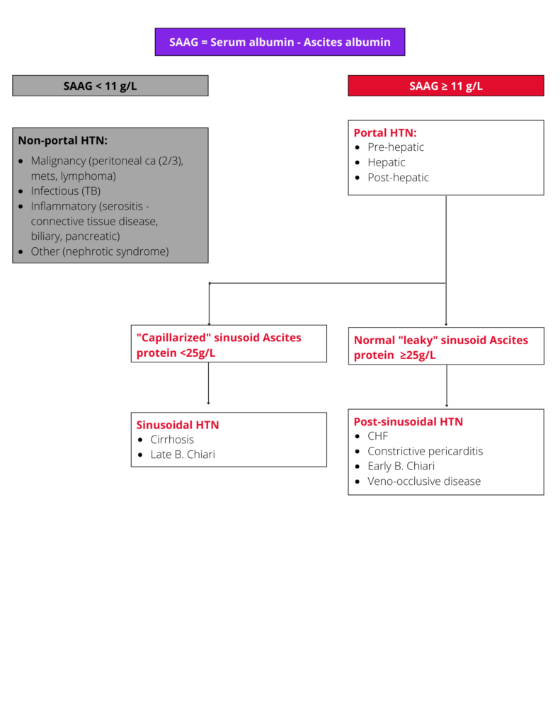
    Serum-ascites albumin gradient

    Substituting Spironolactone with Amiloride

    Amiloride has a weaker diuretic effect but can be substituted for Spironolactone if painful gynecomastia becomes a problem.

    Starting dose of 10mg

    where 10mg = 100mg of Spironolactone

    Maximum dose of 40mg of Amiloride. 

    More information on Spironolactone & Furosemide

    A ratio of 50-100mg Spironolactone to 20-40mg Furosemide often works to balance the electrolytes, but must be individualized.

    The ratio of diuretics can be adjusted to control hyperkalemia (i.e., if hyperkalemia is present, reduce Spironolactone).

    If the electrolytes and creatinine are ok, doses can be increased in increments of 50mg Spironolactone to 20mg Furosemide.

    For example, starting doses of Spironolactone 50mg and Furosemide 20mg can go to 100mg and 40mg respectively after 4-7 days.

    See more information on using the urine sodium

    #1: Dietary non-compliance - Spot urine Na/K ratio is >1 OR the 24-hour urine Na excretion is >78 mEq/day in a patient on diuretics who is not losing weight This means the patient is sensitive to diuretics but not adherent to sodium restriction.
    #2: Diuretic resistance - Spot urine Na/K ratio is ≤1 OR 24-hour urinary Na excretion is <78 mEq per day in a patient on maximally tolerated diuretics who is not losing weight. This means the patient is resistant to diuretics

    Introducing Dr. Abraldes, Dr. Tandon and Michelle Carbonneau

    Video 1 – The top general tips that may be useful for you to know about this page as a family physician

    Video 2 - Transjugular intrahepatic portosystemic shunt (TIPS) for a patient with cirrhosis and ascites or hydrothorax – indications and cautions.

    Patient materials:

    You can direct patients to the following:

    Ascites

    Paracentesis

    Lab tests

    TIPS

    IPC

    Breathing trouble

    Portal hypertension

    References:

    This section was adapted from content using the following evidence based resources in combination with expert consensus. The presented information is not intended to replace the independent medical or professional judgment of physicians or other health care providers in the context of individual clinical circumstances to determine a patient’s care.

    Authors: Dr. Marilyn Zeman, Dr. Guadalupe Garcia-Tsao, Dr. Vijey Selvarajah, Dr. Brian Buchanan, Dr. Puneeta Tandon

    References:

    1. EASL Clinical Practice Guidelines for the management of patients with decompensated cirrhosis. J Hepatol 2018 Aug;69(2):406-460 PMID 29653741
    2. Diagnosis, Evaluation, and Management of Ascites, Spontaneous Bacterial Peritonitis and Hepatorenal Syndrome: 2021 Practice Guidance by the American Association for the Study of Liver Diseases. Hepatology (Baltimore, Md.)74(2), 1014–1048. https://doi.org/10.1002/hep.31884 PMID 33942342
    3.  Advancing Liver Therapeutic Approaches (ALTA) Consortium (2022). North American Practice-Based Recommendations for Transjugular Intrahepatic Portosystemic Shunts in Portal Hypertension. Clinical gastroenterology and hepatology : the official clinical practice journal of the American Gastroenterological Association20(8), 1636–1662.e36. https://doi.org/10.1016/j.cgh.2021.07.018 PMID 34274511

    Last updated November 24, 2022

    Screen for Complications

    Top tips:

    1. In all patients with compensated cirrhosis, assess whether the patient has clinically significant portal hypertension. If they do and there are no contraindications, consider starting carvedilol (or a non-selective beta-blocker) to reduce the risk of decompensation.
    2.  In those patients with compensated cirrhosis who are not already on carvedilol or a non-selective beta-blocker, a fibroscan score of >20 kPA OR a platelet count of <150,000 tells you the patient should be considered for endoscopy. If the fibroscan score is <20 kPA AND platelet count is >150,000 OR the patient is on carvedilol or a non-selective beta-blocker, screening endoscopy is not needed.
    3. Patients with cirrhosis should undergo HCC surveillance with ultrasound and AFP every six months (notably, although by guidelines the AFP is optional, in our experience this is suggested)
    4. Patients who have had a diagnosis of cirrhosis at anytime (regardless of whether they have improved or even cleared their Hepatitis B or C) should continue to undergo HCC surveillance
    Dr. Ramji cartoon
    Dr. fung cartoon
    Dr. Burak cartoon

    Thank you to Dr. Ramji, Dr. Fung, and Dr. Burak for your efforts creating the content on this page. Check out the bottom of the page for a short video from Dr. Fung!

    Screening for liver related complications

    Compensated cirrhosis

    Defined by: the absence of ascites, hepatic encephalopathy, variceal bleeding or jaundice (see more info)

    Decompensated cirrhosis

    Defined by: the presence of ascites, hepatic encephalopathy, variceal bleeding or jaundice (see more info)

    Introducing Dr. Ramji, Dr. Fung and Dr. Burak

    Video 1 - Automating HCC surveillance - information about automatic recall ultrasound surveillance recall programs in Calgary and Edmonton.

    Patient materials:

    You can direct patients to the following:

    HCC

    Liver biopsy

    Lab tests

    Downloadable content:

    You can download these to print or view offline:

    Baveno guidelines 2021

    AASLD Guidance 2018

    EASL Clinical Practice Guidelines

    References:

    This section was adapted from content using the following evidence based resources in combination with expert consensus. The presented information is not intended to replace the independent medical or professional judgment of physicians or other health care providers in the context of individual clinical circumstances to determine a patient’s care.

    Authors: Michelle Carbonneau NP, Dr. Alnoor Ramji, Dr. Puneeta Tandon

    References:

    1. EASL Clinical Practice Guidelines for the management of patients with decompensated cirrhosis. J Hepatol 2018 Aug;69(2):406-460 PMID 29653741
    2. de Franchis, R., Bosch, J., Garcia-Tsao, G., Reiberger, T., Ripoll, C., & Baveno VII Faculty (2022). Baveno VII – Renewing consensus in portal hypertension. Journal of hepatology76(4), 959–974. https://doi.org/10.1016/j.jhep.2021.12.022 PMID 35120736
    3. EASL Clinical Practice Guidelines: Management of hepatocellular carcinoma. J Hepatol. 2018 Jul;69(1):182-236 PMID 29628281
    4. Loomba R et al. AGA Clinical Practice Update on Screening and Surveillance for Hepatocellular Carcinoma in Patients with Nonalcoholic Fatty Liver Disease: Expert Review. Gastroenterology 2020 Jan 29 PMID 32006545

    Last reviewed November 3, 2022

    Etiology management specific to cirrhosis

    Top tips:

    1. Disease-specific treatment should be considered for all patients with compensated cirrhosis
    2. The decision to treat the disease etiology in patients with decompensated disease needs to be individualized based on the disease and patient factors, and in many cases requires consultation with a liver specialist.
    3. Patient with decompensated cirrhosis should be considered for referral for liver transplant evaluation.
    4. All patients with cirrhosis require Q 6 monthly ultrasound surveillance for hepatocellular carcinoma, even in those with Hepatitis C virus who have cleared the infection.
    Dr. Shah cartoon
    Dr. Abraldes cartoon-needs white hair

    Thank you to Dr. Shah and Dr. Abraldes for your efforts creating the content on this page!

    Hepatitis C

    • Some Hepatitis C patients can now be treated by Primary Care (after seeking advice from hepatology, infectious diseases or gastroenterology – as required by insurance provider). See the following link to the Alberta Health Services Hepatitis C Virus Primary Care Pathway
    • Successful treatment is associated with reductions in morbidity, all-cause and liver-related mortality and an improved quality of life in the majority of patients.
    • Historically, many sub-populations of patients required special management protocols, but that is no longer the case. Careful attention to co-morbidities (especially active Hepatitis B), drug-drug interactions and determining the presence or absence of decompensated cirrhosis should now be the focus of a pre-treatment clinical assessment.
    • Suggested treatment regimens change regularly depending upon the available evidence. The most widely used pangenotypic agents are safe, well tolerated and highly efficacious. See infectiousdiseaseadvisor.com OR hcvguidelines.org for a link to suggested treatment regimens and associated evidence.
    • Special treatment considerations in patients with decompensated cirrhosis
      • Patients with a MELD score of >20 and/or a GFR of <30 who are candidates for liver transplantation should have treatment decisions supervised by a transplant center. In most cases these patients should be treated after transplant to optimize the chance of viral clearance
      • Protease-inhibitor based regimens including Maviret® (glecaprevir and pibrentasvir) and Vosevi® (sofosbuvir, velpatasvir, voxilaprevir) are contraindicated.

    Non alcoholic fatty liver disease (NAFLD)

    • The mainstays of NAFLD treatment are optimal management of metabolic disease, weight loss, dietary support, statin use when appropriate (statins are not contraindicated), and liver-fat specific interventions when evidence suggests benefit.
    • Aim for a dietitian supervised 7-10% weight loss using a 500-1000 kcal energy deficit. A Mediterranean style diet has been associated with reductions in liver fat. Processed foods and fructose-containing beverages and foods should be avoided.
    • Patients with cirrhosis and NAFLD are at risk of muscle mass loss if weight loss is carried out without appropriate dietary counselling and concurrent increases in physical activity.
    • Patients should be encouraged to participate in 150 minutes per week of moderate intensity activity (aerobic + resistance training). Both of these modalities reduce steatosis.
    • Statins may be used to reduce LDL-cholesterol and prevent cardiovascular risk. Of note, with worsening liver function, there is an increase in a membrane transporter in the hepatocytes that modulates statin uptake (SLCO1B), resulting in higher serum statin levels and increased rates of myopathy and hepatotoxicity. See Child Pugh calculator link here.
      • In patients with Child Pugh A disease, statins CAN BE USED at regular doses.
      • In patients with Child Pugh B disease, one agent has the most evidence to support safety – Rosuvastatin at a dose of 5 mg. Monitor CK levels and symptoms to detect adverse events.
      • In patients with Child Pugh C disease, statins are unlikely to have benefit and very likely to cause harm – they should be DISCONTINUED.
    • Atorvastatin should NOT be used in cirrhosis
    • Vitamin E can be considered at a dose of 400 IU/day in select patients without cardiovascular disease
    • Bariatric surgery can be considered in selected patients (BMI >35) with compensated cirrhosis. In the setting of decompensated cirrhosis or portal hypertension, the risks are very high and surgery should not be considered until at or after transplant.
    • There are many novel agents under investigation that act along the entire pathway that contributes to steatohepatitis.

    Hemochromatosis

    • Can occur as a primary or secondary condition. The most common cause of primary hemochromatosis is an alteration in the HFE gene, but other genetic mutations can also lead to primary disease.
    • In patients of European ancestry, the most common cause of hemochromatosis is a mutation in the HFE gene.
        • Among individuals of European ancestry, the carrier frequency of the most significant mutation (C282Y) is ~1/10.
        • Clinical manifestations of iron overload can be variable even in patients with C282Y homozygosity, as there is incomplete penetrance.
    • The goal of treatment is to prevent organ dysfunction and improve organ function by reducing excess iron. All contributing factors (i.e. alcohol use, HCV infection) should be screened for and addressed as these can increase iron stores.
    • Phlebotomy is the mainstay of therapy for patients with iron overload. It is associated with improvements in survival, hepatic fibrosis (with reduction in the size of varices), cardiomyopathy and hypogonadism. It does not improve joint pain.
    • Phlebotomy schedules can be tailored to the individual, and can be done weekly to every two weeks if tolerated (hemoglobin >110 g/dL, hemodynamic stability). Each unit of blood removes 200-250 mg of iron. Target ferritin values of 50-100 ng/mL. Once this value is reached, maintenance is required at approximately 1 unit of blood very 2-4 months. Of note, patients with cirrhosis are more likely to become anemic with phlebotomy.
    • Patients who do not tolerate phlebotomy should be considered for chelation therapies.
    • Q 3 monthly labs to check CBC, ferritin, iron saturation, reticulocyte count
    • Dietary restriction of iron is not required. Patients should avoid supplements that contain additional iron. Patients should avoid vitamin C supplements. Uncooked seafood should be avoided given the risk of vibrio vulnificus infection (a bacteria that thrives on iron).
    • Relatives of any person with genetic hemochromatosis should be considered for testing, usually in a sequential pattern.

    Hepatitis B

    • Decision-making around Hepatitis B treatment is based upon considering the individual’s hepatitis B status, degree of liver fibrosis, and projected natural history with and without treatment. It is important to recognize there is a limitation in the evidence base around the optimal time to start and stop treatment so collaborative decision making is very important.
    • Scenarios where treatment initiation is widely accepted include: patients with active disease who are developing progressive fibrosis, patients with advanced liver disease especially decompensation, patients with high viral load where the goal is to prevent vertical transmission, patients about to undergo immunosuppression, and patients who develop hepatocellular cancer.
    • Note, in the setting of acute on chronic liver failure or decompensated cirrhosis with a detectable HBV DNA level, antiviral therapy should also be started as it results in clinical and biochemical improvement in 40-50% of cases. ~ 1/3 of these patients can be delisted for liver transplantation.
    • The most commonly used treatments in cirrhosis are Entecavir, Tenofovir disoproxil fumarate (TDF) or Tenofovir alafenamide (TAF). Tenofovir should be avoided with renal dysfunction. There have been rare reports of lactic acidosis in patients with severe liver dysfunction who are started on Entecavir.
    • In patients on treatment, monitor HBV DNA and ALT levels every 3 months until aviremia is achieved. Then testing can be extended to every 6 monthly. Patients on Tenofovir also require q 3-6 monthly monitoring of renal function and serum phosphate levels.
    • Patients with cirrhosis or HCC should continue to receive treatment indefinitely or until HBsAg loss.

    Autoimmune hepatitis

    • The management of autoimmune hepatitis is best performed in a specialized hepatology clinic.
    • Initial therapy requires the assessment for potential contraindications to therapy (Prednisone, Azathioprine) especially evaluation for any active infection.
    • It is most common to give a combination of Prednisone and Azathioprine in the early phases of management:
      • Start with Prednisone alone x 2 weeks and then add on Azathioprine starting at week 3.
      • Prednisone dose is 0.5 mg/kg body weight/day tapering down by 10 mg per week until at 20 mg and then by 2.5-5 mg every 2-4 weeks until off.
      • Azathioprine dose starts at 50 mg per day for weeks 3 and 4 (provided the serum bilirubin is below 100 umol/L) increasing to 1-2 mg/kg body weight per day by week 5 if tolerated.
    • Maintenance therapy can consist of a single agent or a combination of immunosuppressives.

    Primary biliary cholangitis

    • All patients should receive ursodeoxycholic acid (UDCA) at a dose of 13-15 mg/day (either as a single daily dose or in divided doses if there are concerns about tolerability.
    • UDCA is generally well tolerated with minimal side effects (rarely weight gain, hair thinning, diarrhea, flatulence have been reported).
    • Patients should continue UDCA for life, even in the absence of biochemical improvements, as this is associated with prolonged LT-free survival.
    • Biochemical response should be assessed at 1 year with lack of response defined by the ALP >1.67 x ULN and/or bilirubin >1.0 x ULN.
    • Risk factors for an inadequate biochemical response are early age at diagnosis (<45 years), male sex, and advanced fibrosis at presentation.
    • Patients with a lack of response to UDCA require the addition of second line therapy including obeticholic acid (licensed) and bezafibrate (off-label).
      • Adjunctive treatment for incomplete responders consists of obeticholic acid (OCA) as the first option (use only in Child Pugh A disease), with bezafibrate (400 mg daily) and clinical trials as alternatives.
      • Obeticholic acid can be dosed at 5 mg po daily increasing to 10 mg po daily at six months depending upon tolerance.  Side effects include pruritus and elevations in the serum cholesterol.
      • Bezafibrate is dosed at 400mg daily. Side effects include GI upset and increased ALT.
    • Patients should be monitored q 6-12 monthly with bilirubin, ALP, AST, albumin, platelet count and transient elastography.
    • Patients should be evaluated for associated disease related symptoms and complications (pruritus, sicca complex, fatigue, osteoporosis, fat soluble vitamin deficiency)
    • Prognosis can be estimated using risk scores such as the GLOBE score (https://www.globalpbc.com/globe). Patients with risk scores >0.30 have significantly shorter transplant-free survival than matched healthy individuals (P < .0001).

    Primary sclerosing cholangitis

    • The role of ursodeoxycholic acid (UDCA) remains unclear.
      • It is known that high dose (20-25mg/kg/d) UDCA is harmful. A UDCA trial for 6-12 months at a low dose (13-15 mg/kg per day in divided doses) can be considered with continuation of therapy if the ALP decreases by at least 40% or there is symptomatic improvement.
    • The role of cholangiocarcinoma screening remains unclear. Notably, many centers carry out annual screening with ultrasound or MRCP ± measurement of a serum CA 19-9 (Bowlus CL et al. 2019). Given the lack of data it is reasonable to forego routine screening. If a dominant stricture is suspected, the patient should be referred to a tertiary care center with expertise in investigating these cases.
    • All patients with PSC should have an annual ultrasound to screen for gallbladder carcinoma (this will be captured in q 6 monthly HCC surveillance in patients with cirrhosis).
    • 90% of patients with PSC have IBD. If colitis has been established, surveillance colonoscopy is required q 1-2 yearly. Those patients without IBD require colonoscopy q 3-5 yearly with biopsies to look for previously undiagnosed colitis.
    • bone density evaluation should be carried out at diagnosis and q 2-3 yearly
    • Prognosis can be estimated using risk scores such as the PRESTO (https://rtools.mayo.edu/PRESTO_calculator/)

    Calculators:

    Use these calculators:

    GLOBE PBC prognostic score

    PRESTO PSC prognostic score

    References:

    This section was adapted from content using the following evidence-based resources in combination with expert consensus. The presented information is not intended to replace the independent medical or professional judgment of physicians or other health care providers in the context of individual clinical circumstances to determine a patient’s care.

    Authors: Dr. Aldo Montano-Loza, Dr. Hemant Shah, Dr. Carla Coffin, Dr. Puneeta Tandon

    References:

    1. Mack CL et al. Diagnosis and management of autoimmune hepatitis in adults and children: 2019 practice guidance and guidelines from the American Association for the study of liver diseases. Hepatology 2019. PMID 31863477
    2. Kowdley K et al. ACG Clinical Guideline: Hereditary Hemochromatosis. The American Journal of Gastroenterology. August 2019. PMID 31335359
    3. Ghany M et al. Hepatitis C Guidance 2019 Update: AASLD-IDSA Recommendations for Testing, Managing, and Treating Hepatitis C Virus Infection. Hepatology 2019. PMID 31816111
    4. Terrault NA et al. Update on Prevention, Diagnosis, and Treatment of Chronic Hepatitis B: AASLD 2018 Hepatitis B Guidance. Hepatology 2018. PMID 29405329
    5. EASL-EASD-EASO Clinical Practice Guidelines for the management of non-alcoholic fatty liver disease. Journal of Hepatology 2016. PMID 27055256
    6. Lindor KD et al. Primary Biliary Cholangitis: 2018 Practice Guidance from the American Association for the Study of Liver Diseases. Hepatology 2018. PMID 30070375
    7. Corpechot C, et al. Noninvasive elastography-based assessment of liver fibrosis progression and prognosis in primary biliary cirrhosis. Hepatology 2012. PMID 22271046
    8. EASL Clinical Practice Guidelines: The diagnosis and management of patients with primary biliary cholangitis. J Hepatol 2017. PMID 28427765
    9. Bowlus CL, Lim JK, Lindor KD. AGA Clinical Practice Update on Surveillance for Hepatobiliary Cancers in Patients With Primary Sclerosing Cholangitis: Expert Review. Clin Gastroenterol Hepatol 2019. PMID 31306801
    10. Pose E et al. Safety of two different doses of simvastatin plus rifaximin in decompensated cirrhosis (LIVERHOPE-SAFETY): a randomized, double-blind, placebo-controlled, phase 2 trial. Lancet Gastroenterol Hepatol 2020 Jan;5(1):31-41. PMID 31607677
    11. Pape S et al. Predniso(lo)ne Dosage and Chance of Remission in Patients with Autoimmune Hepatitis. Clin Gastroenterol Hepatol 2019 Sep;17(10):2068-2075. PMID 30625402
    12. Sanyal A et al. Pioglitazone, vitamin E, or placebo for nonalcoholic steatohepatitis. N Eng J Med 2010 May;6(362):1675-1685. PMID 20427778
      List of authors.

    Last reviewed December 9, 2022. 

    Work up the etiology

    Top tips:

    1. The three most common causes of cirrhosis are Non-alcoholic fatty liver disease, Alcohol and Hepatitis C virus
    2. In addition to a detailed history, examination and liver imaging such as an ultrasound doppler, basic testing in all patients with cirrhosis should include screening with the anti-HCV antibody (will require HCV PCR to confirm diagnosis if antibody is positive), HBsAg, quantitative immunoglobulins and iron studies

    Order panels Liver disease work up

    For adults:
    Liver disease work up 

    Dr. Zeman cartoon

    Check out the bottom of the page for a short video from Dr. Zeman!

    Cirrhosis Etiologies

    Most common:

    • Nonalcoholic Fatty Liver Disease (NAFLD)
    • Alcohol related liver disease
    • Chronic viral hepatitis (Hepatitis B, C)

    Less common causes (not all inclusive):

    • Autoimmune hepatitis
    • Hereditary Hemochromatosis
    • Primary biliary cholangitis (PBC)
    • Primary sclerosing cholangitis (PSC)
    • Wilsons disease
    • Alpha-1 antitrypsin deficiency

    Etiology Considerations

    History:

    • Alcohol: Alcohol use (>2 standard drinks/day for women and >3 for men where a standard drink is 10-14 grams of alcohol)
    • Non-alcoholic fatty liver disease: Diabetes or metabolic syndrome
    • Viral hepatitis: Exposures (place of birth, travel, sexual behaviours, drug use, incarceration)
    • Medications and supplement use are unlikely to cause cirrhosis, but can contribute to acute deterioration – see link to Livertox website to evaluate risk and injury pattern of high-risk medications or medications started within the previous 6 months.
    • Autoimmune hepatitis, Primary biliary cholangitis: Family history or personal history of other autoimmune conditions
    • Primary sclerosing cholangitis: Co-existing Inflammatory bowel disease
    • Genetic liver diseases (Wilsons, Alpha-1-antitrypsin deficiency, Hemochromatosis): Family history of cirrhosis (age of onset, etiology), hemochromatosis, premature emphysema
    • History of congestive heart disease

    Pattern of liver tests:

    • Hepatocellular (Predominantly high AST/ALT compared to ALP)
    • Cholestatic (Predominantly high ALP compared to AST/ALT)

    Link to R-Factor for Liver Injury calculator when you are working up worsening liver enzymes to determine if the pattern is hepatocellular, cholestatic or mixed.

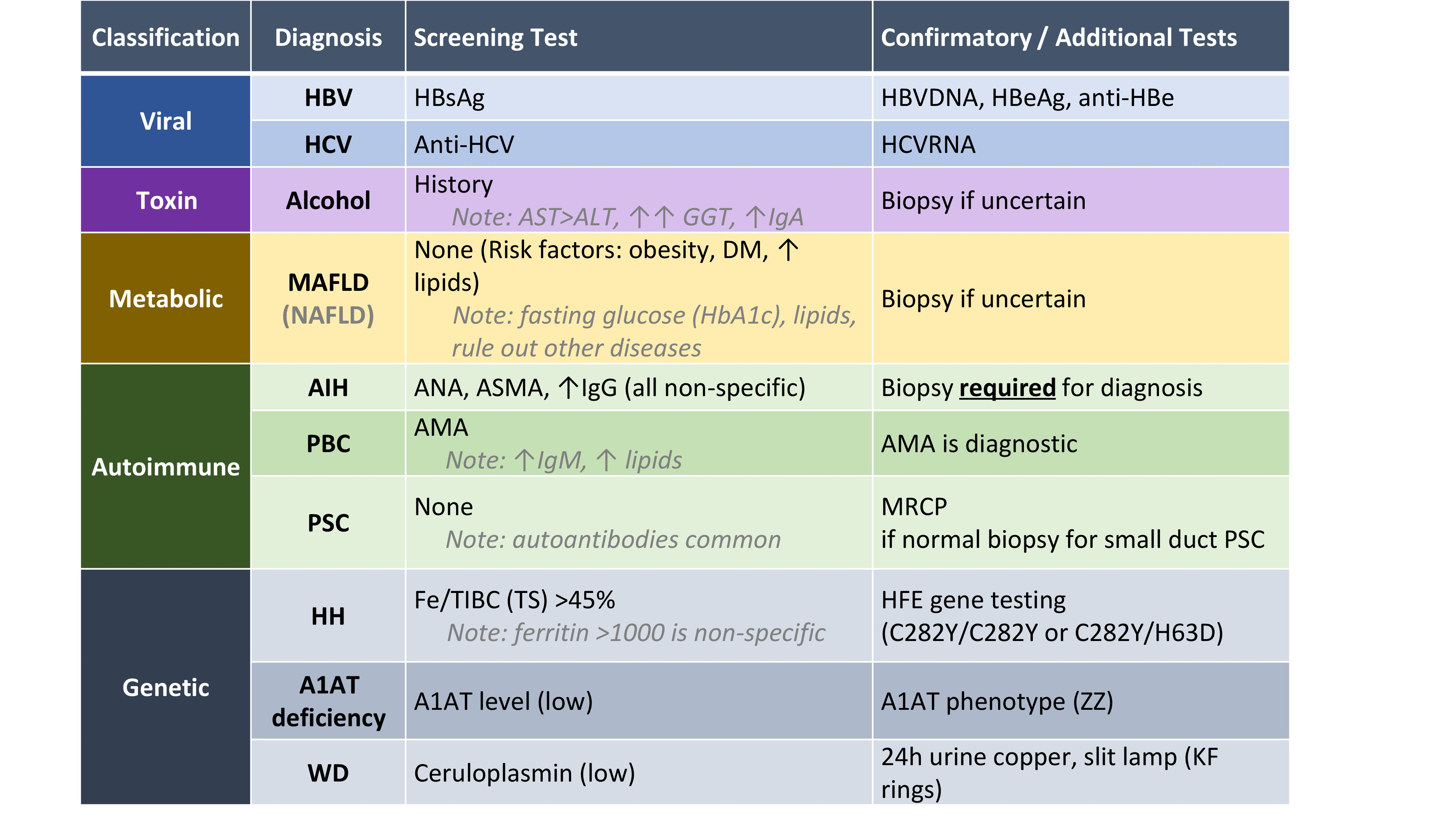
    Initial Lab investigations

    In addition to a detailed history and relevant physical examination, the following laboratory investigations can be done to determine the etiology in cases of confirmed cirrhosis (image courtesy of Dr. Kelly Burak):

     Introducing Dr. Zeman

    Video 1 - The top tips that may be useful for you to know about this page as a family physician including: Investigations to order while waiting for a specialist appointment; and When you need to call a specialist more urgently.

    References:

    This section was adapted from content using the following evidence based resources in combination with expert consensus. The presented information is not intended to replace the independent medical or professional judgment of physicians or other health care providers in the context of individual clinical circumstances to determine a patient’s care.

    Authors: Michelle Carbonneau NP, Dr. Marilyn Zeman, Dr. Aldo Montano-Loza, Dr. Kelly Burak, Dr. Puneeta Tandon

    Last reviewed December 9, 2022

    Making a diagnosis of cirrhosis (+ a link to the AHS Fatty Liver pathway)

    Top tips:

    1. In patients with risk factors for chronic liver disease AST, ALT and platelet count can help to rule out cirrhosis.
    2. A FIB 4 (calculated using age, AST, ALT and platelet count) cut-off score of < 1.3 is a good predictor of the ABSENCE of cirrhosis in all clinical contexts.
    3. As per the Alberta Health Services Fatty liver pathway (link), if your patient has a FIB-4 of more than or equal to 1.3, refer to a specialist trained in the management of liver disease.
    4. Elastography is a good tool to assess liver fibrosis. Normal results of elastography can help to rule out cirrhosis and values over 15 kPa (by transient elastography) strongly suggest cirrhosis.
    5. A low platelet count (<150 G/L) is an important clue that a patient may have cirrhosis.
    6. Patients with suspicion of cirrhosis based on the above-mentioned tests should be referred to a liver clinic. Liver specialists will use clinical tools as well as a non-invasive approach based on liver Elastography and abdominal ultrasound to identify cirrhosis.
    7. In unclear cases, invasive tests (liver biopsy, hepatic venous pressure gradient measurement, endoscopy) will be required to come to a final diagnosis.
    Dr. Abraldes cartoon-needs white hair
    Dr. Berzigotti cartoon

    Check out the bottom of the page for short videos from Dr. Abraldes and Dr. Berzigotti!

    Physical exam findings
    Spider Nevus

    Images courtesy of Patrick Kamath, MD

    • Dilated arterioles characterized by prominent central arteriole and radiating vessels, usually found in distribution of superior vena cava
    • Compression of central arteriole causes blanching of the “spider”
    • More than 3 spider nevi is considered abnormal


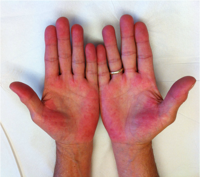
    Palmar Erythema

    Image courtesy of Patrick Kamath, MD

    • Intense red coloration of the thenar and hypothenar eminence
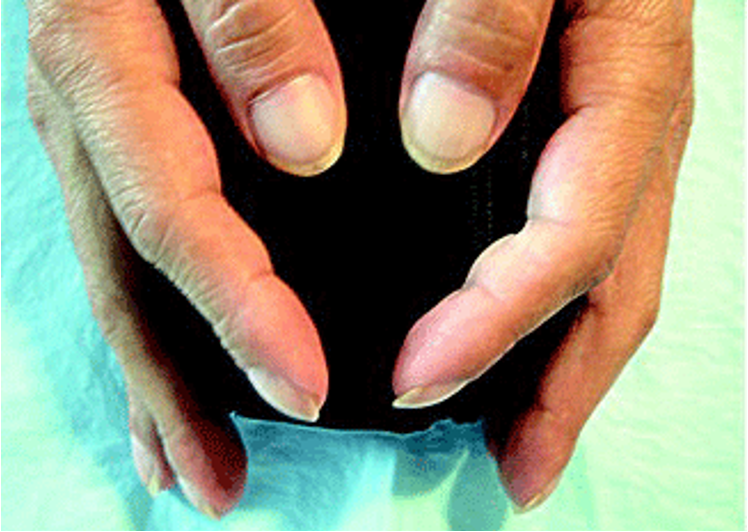
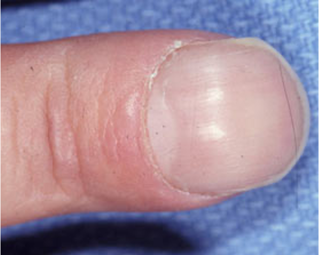
    Terry Nails


    Image courtesy of Patrick Kamath, MD

    • Characterized by proximal nail bed pallor
    • Can also involve the entire nail plate
    • Predominant involvement of the thumb and index finger
    Clubbing

    Image courtesy of Patrick Kamath, MD

    • Associated with hepatopulmonary syndrome
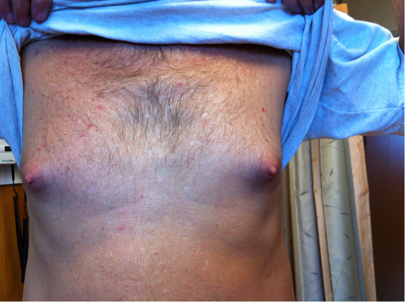
    Gynecomastia

    Image courtesy of Patrick Kamath, MD

    • Enlargement of male breasts with palpable tissue
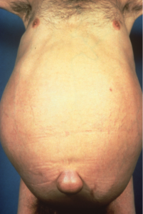
    Caput Medusae and Ascites


    Image courtesy of Patrick Kamath, MD

    Ultrasound findings

    Image courtesy of Dr. A Berzigotti

    Figure summarizing the most common ultrasound findings in patients with cirrhosis


    A) Nodular liver surface (arrows)

    B) Liver vein with a reduced diameter and irregular walls due to the compression of the surrounding cirrhotic parenchyma. Heterogeneous parenchyma

    C) Hepatofugal (away from the liver) blood flow in the portal vein. This finding suggests very high resistance in the liver and lower resistance outside the liver, usually due to the presence of large porto-systemic collaterals

    D) Left intrahepatic branch of the portal vein shows hepatofugal blood flow

    E) 2D-Shear Wave Elastography shows very high liver stiffness values (> 55 kPa). Small amount of ascites around the liver surface

    F) Enlargement of the spleen quantified by ultrasound by measuring the pole to pole distance. Here is 16 cm (severe splenomegaly)

    G) Porto-systemic collaterals close to the spleen appear as large anechoic channels

    H) By using color-Doppler, the channels show venous flow in the direction of the renal vein, confirming the diagnosis of spleno-renal collaterals

    I) In the left lobe (sagittal view) a long channel can be often observed between the left portal vein and the extrahepatic space, within the ligamentum rotondum. This corresponds to a patent paraumbilical vein

    L) The diagnosis can be confirmed by using color-Doppler, which shows hepatofugal flow within this vessel

    M) Advanced cirrhosis: small liver, with nodular surface and heterogeneous parenchyma. The portal vein is dilated, and 50% of the lumen is occupied by echogenic material, confirming the diagnosis of portal vein thrombosis. The liver is surrounded by ascites

    Quality considerations for Transient Elastography

    Patient should be fasting for at least 2 hours

    Quality criteria:

    • Need 10 Valid shots
    • Need >60% success rate = valid shots/total shots
    • Need IQR (Interquartile range)/M (median liver stiffness measurements) ≤ 0.3

    Consider the following parameters that may impact interpretation:

    • Serum aminotransferases ≥ 5 x ULN
    • BMI (use XL probe if BMI >30 kg/m2 or skin-to-capsule distance >25 mm)
    • Extra-hepatic cholestasis
    • Right heart failure or other causes of congestive liver
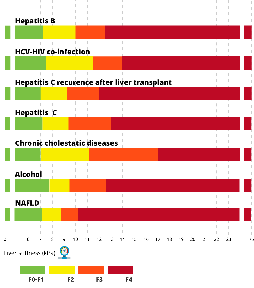
    Transient Elastography cut-offs

    Liver biopsy findings
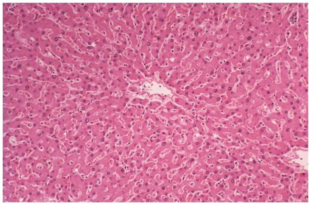
    Normal

    Image courtesy of Patrick Kamath, MD

    In the normal liver, plates of hepatocytes are arranged in a concentric fashion around the portal tract (blue curved arrow)


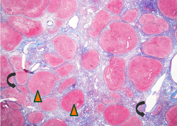
    Cirrhosis

    Image courtesy of Patrick Kamath, MD

    In cirrhosis, the normal hepatic architecture is replaced by diffuse fibrosis (black curved arrows) with nodular regeneration (arrowheads)

    Introducing Dr. Abraldes and Dr. Berzigotti

    Video 1 - The top tips that may be useful for you to know about this page as a family physician including: How the page fits with the Alberta fatty liver pathway ; Calculating Fib-4 ; and When you should refer!

    Video 2 - The clues Dr. Berzigotti uses in her practice to make a diagnosis of cirrhosis.

    Video 3 - A quick tour of the ultrasound findings of cirrhosis.

    Video 4 - Practical tips for performing transient elastography more accurately.

    Patient materials:

    You can direct patients to the following:
    What the liver does

    What is cirrhosis?

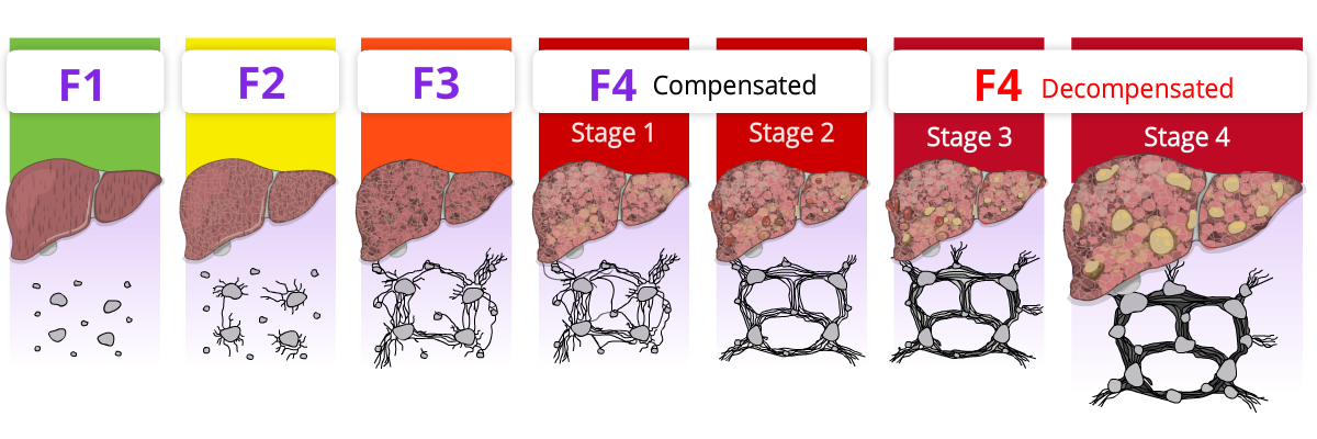
    Stages of cirrhosis

    Living with cirrhosis

    : Links and Calculators

    Alberta Health Services link to the fatty liver pathway

    Use these calculators to help with the diagnosis:
    APRI

    Fib 4

    NAFLD score

    Child Pugh

    MELD-Na

    Downloadable content:

    You can download these to print or view offline:
    EASL Guidelines

    References:

    This section was adapted from content using the following evidence based resources in combination with expert consensus. The presented information is not intended to replace the independent medical or professional judgment of physicians or other health care providers in the context of individual clinical circumstances to determine a patient’s care.

    Authors: Dr. Mang Ma, Dr. Annalisa Berzigotti, Dr. Puneeta Tandon

    References:

    1. EASL-ALEH Clinical Practice Guidelines: Non-invasive tests for evaluation of liver disease severity and prognosis. J Hepatol 2015 Jul;63(1):237-64 PMID 25911335
    2. Kelly EMM et al. An Assessment of the Clinical Accuracy of Ultrasound in Diagnosing Cirrhosis in the Absence of Portal Hypertension. Gastroenterology and Hepatology 2018 Jun; 14(6):367-373 PMID 30166950

    Last updated November 10, 2022.